
For the 3D printing of organ models which contains the live cells researchers at the University of Buffalo have come up with a fine-tuned use of stereolithography. The technique developed by the team is capable of printing the models 10 to 50 times faster than what the industry standard is capable, as described in the journal Advanced Healthcare Materials. This marks as a major step in the quest for the creation of 3D-printed replacement organs.
Traditional 3D printing requires the careful addition of material to the 3D model with the help of small needle which can result in fine details but the process is very slow. It takes six to seven hours to print the model of any human part, for example the hand. Cellular stress and injury take place due to the lengthy process; this inhibits the ability to seed the tissues with live, functioning cells.
The method adopted by the Buffalo team helps in decreasing the damage to live cells. This rapid, cell-friendly technique is an important step towards the path of creating printed tissues which will be infused with large numbers of living cells, this technique is funded by US National Institute of Biomedical Imaging and Bioengineering (NIBIB).
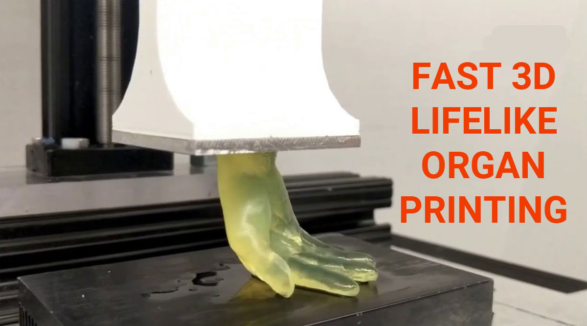
Through this technique developed by the team, there will be no need to use a needle which will move across a surface slowly to build the details of the 3D model. A system has been developed by the team which allows a small 10 cm model of a human hand to rise out of a liquid mass in very less time. What happens in this stereolithographic method is that it uses projected light at the bottom of the tank to be able to penetrate right through the mix of hydrogel and live cells. The light in this process polymerises the hydrogel/cell mixture at the accurately right positions in the model, thereby building entire layers of the model in a continuous manner, instead of one pinpoint at a time.
The Director of the NIBIB program in Synthetic Biological Systems, Dr David Rampulla has said “This is a significant step towards the printing of biologically active 3D tissues,” he also added “This new technique combines a hydrogel mix that is very cell friendly with the rapid printing process, which spares cells from being suspended in a cell-damaging environment for an extended period. The combination has allowed the team to introduce live cells into their 3D-printed tissues, with the vast majority of cells remaining alive and functional.”
The aim is to move from small models of organs to full-sized replacement organs in the near future. This new method has been successfully used by the engineering team to print tissues which will contain inner branching networks that can mimic blood vessels. The Associate Professor Rougang Zhao who is the leader of the team has been noted saying, “To keep alive cells that are deep inside a full-sized printed organ is one of the many challenges in creating functional replacement organs. We found that our method performed well in terms of creating branched, vessel-like networks to facilitate delivery of nutrients to cells embedded throughout the printed tissue.”
The team has also gone a step further to introduce live endothelial cells into the branching canals of their 3D-printed tissues. In the walls of the artificial vasculature the endothelial cells stick to and expands to form an endothelial lining which is similar to the ones found in actual blood vessels. Currently there is an ongoing work to increase the size of the printed tissues while at the same time maintaining structural integrity and cell viability by the team. The work by the team is an important step towards the goal of being able to print replacement organs one day. This advancement in biomedical can be helpful to save countless number of lives that are lost due to the shortage of donor organs.